Leg Bones Diagram / Figure 12-33. Human anatomy diagrams show internal. Its lower end helps create the knee joint. The femur in the thigh; The largest and most medial leg bone, forming both the knee and ankle joints. Here are a few anatomical plates about the leg and the foot.
Learn how to draw the femur, patella, tibia, and fibula in this lesson! The foot bones shown in this diagram are the talus, navicular, cuneiform, cuboid, metatarsals. The foot bones shown in this diagram are the talus, navicular, cuneiform, cuboid. The femur in the thigh; Your leg bones are the longest and strongest bones in your body.
An Introduction to Skeletal System - The Bones and What They Do from www.exploringnature.org Bone structure diagram wiring diagrams click. The largest and most medial leg bone, forming both the knee and ankle joints. Health diagram bone skeleton leg knee science anchor chart human human body. The patella in the knee; The knee joint is the largest joint in the body and is primarily a hinge joint although some sliding and rotation. Nervsystemet anatomy, diagram & function | health. The femur, or thighbone, is the longest and largest bone in the human body. Bones of the leg and foot, lower leg bone anatomy, leg bones anatomy, leg muscles, leg bones diagram, leg bone structure, leg anatomy muscles, parts of the lower leg.
Visit kenhub for more skeletal system quizzes.
The bones of the leg are the femur, tibia, fibula and patella. He leg's main function in the human is for locomotion and support of the rest of the body. The knee joint is the largest joint in the body and is primarily a hinge joint although some sliding and rotation. Bone anatomy of knee joint. The human leg consists of 8 bones, 4 per leg. Learn how to draw the femur, patella, tibia, and fibula in this lesson! The foot bones shown in this diagram are the talus, navicular, cuneiform, cuboid, metatarsals. You'll learn about the muscles, bones, and other structures of each area of the leg. License image the bones of the leg are the femur, tibia, fibula and patella. Diagram and names of leg bones, diagram of foot and leg bones, diagram of leg bones, diagram of lower leg bones related posts of diagram of leg bones. Your legs are two of your most important body parts. When you stand or walk, all the weight of your upper body rests on them. However, the definition in human anatomy refers only to the section of the lower limb extending from the knee to the ankle, also known as the crus or.
Knee bone diagram illustrations & vectors. Time to jump right into the biggest and strongest bones in the human body. The patella in the knee; License image the bones of the leg are the femur, tibia, fibula and patella. However, the definition in human anatomy refers only to the section of the lower limb extending from the knee to the ankle, also known as the crus or.
Lab 1 at University of North Carolina-Wilmington - StudyBlue from s3.amazonaws.com Leg femur diagram data wiring diagram today. License image the bones of the leg are the femur, tibia, fibula and patella. Ankle and foot pain massage therapy connections. The patella in the knee; Knee bone diagram illustrations & vectors. This page is about leg bones diagram,contains aluminium plant safety: The foot bones shown in this diagram are the talus, navicular, cuneiform, cuboid. Your leg bones are very large and strong to help support the weight of your body.
Ankle and foot pain massage therapy connections.
Your leg bones are very large and strong to help support the weight of your body. Learn how to draw the femur, patella, tibia, and fibula in this lesson! When you stand or walk, all the weight of your upper body rests on them. Use the leg bones diagrams to learn the names of the leg bones. Here are a few anatomical plates about the leg and the foot. Quizzes on human skeletal system anatomy, bone anatomy, and bone markings. It mainly serves as an attachment point for the muscles of the lower leg. Most relevant best selling latest uploads. He leg's main function in the human is for locomotion and support of the rest of the body. Bone structure diagram wiring diagrams click. Human anatomy diagrams show internal. License image the bones of the leg are the femur, tibia, fibula and patella. Its lower end helps create the knee joint.
The human leg consists of 8 bones, 4 per leg. Bone structure diagram wiring diagrams click. Here are a few anatomical plates about the leg and the foot. The foot bones shown in this diagram are the talus, navicular, cuneiform, cuboid, metatarsals and calcaneus. The human leg, in the general word sense, is the entire lower limb of the human body, including the foot, thigh and even the hip or gluteal region.
Labeled Skeletal System Diagram | Names, The o'jays and Medical from s-media-cache-ak0.pinimg.com Here are a few anatomical plates about the leg and the foot. The foot bones shown in this diagram are the talus, navicular, cuneiform, cuboid, metatarsals. At the same time, the bones and joints of the leg and foot must be strong enough to support the body's weight while remaining flexible enough for movement and balance. The femur, or thighbone, is the longest and largest bone in the human body. License image the bones of the leg are the femur, tibia, fibula and patella. Click now to learn more about the bones, muscles, and soft tissues tibia: The knee joint is the largest joint in the body and is primarily a hinge joint although some sliding and rotation. Learn how to draw the femur, patella, tibia, and fibula in this lesson!
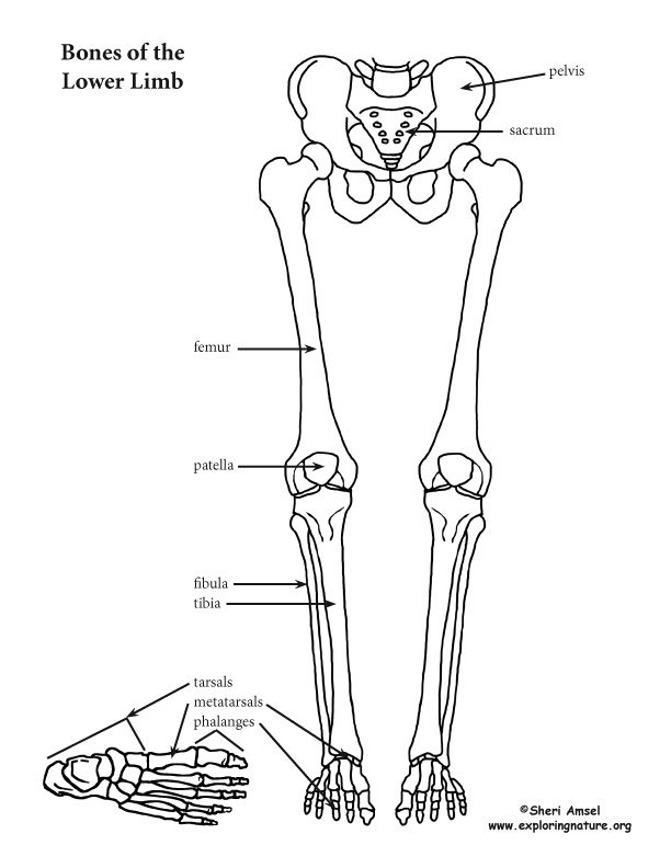
These simple labelled diagrams of the bones of the lower legs and feet and the bones of the arms and hands this diagram shows the skeletal structure of the leg (anterior view) and foot (dorsal view).
The bones of the leg are the femur, tibia, fibula and patella. Most relevant best selling latest uploads. Bones of the leg and foot, lower leg bone anatomy, leg bones anatomy, leg muscles, leg bones diagram, leg bone structure, leg anatomy muscles, parts of the lower leg. The bones involved in it, however, are only the femur and the tibia, although the smaller bone of the leg, the fibula, is carried along in the movements of flexion, extension, and slight rotation that this joint. The femur, or thighbone, is the longest and largest bone in the human body. Bone diagram barca fontanacountryinn com. In the leg, the interosseous membrane extends between the tibia and the fibula, running along the crests of the bones. Knee bone diagram illustrations & vectors. This page is about leg bones diagram,contains aluminium plant safety: He leg's main function in the human is for locomotion and support of the rest of the body. Nervsystemet anatomy, diagram & function | health. Bone anatomy of knee joint. The femur in the thigh;
Share :
Post a Comment
for "Leg Bones Diagram / Figure 12-33"


Post a Comment for "Leg Bones Diagram / Figure 12-33"